Sunday, 4 December 2016

mTRAP: Gene knockouts trap malaria in the erythrocyte

Malarial TRAP family proteins were thought to function during cell invasion, with the merozoite form MTRAP involved in asexual erythrocytic infection. However, a recent study of gene disruption using knockouts and the CRISPR-Cas9 system reveal unexpected roles for the MTRAP protein in cell egress and gamete development. 

The TRAP (thrombospondin-related anonymous protein) family are conserved in various apicomplexan parasites causing severe human disease, including malaria (Plasmodium spp), and toxoplasmosis (Toxoplasma spp). Previous research studying the TRAP protein family had suggested functions during cell invasion and gliding motility 2. Whilst this may hold true for some of the protein family members, research undertaken by Bargieri et al 1 has revealed the merozoite TRAP protein (MTRAP) operates in an unexpected way.

Using gene knockouts in the malaria animal model Plasmodium berghei, and the CRISPR-Cas9 system in human Plasmodium falciparum, the research suggests that MTRAP deficiency results in a transmission block. Without MTRAP, neither male nor female gametocytes are able to egress from erythrocytes in the mosquito, which prevents zygote formation and further transmission. It had been thought MTRAP was expressed in the Plasmodium asexual merozoite, responsible for the erythrocytic cycle, where it mediated adhesion and invasion of the red blood cell. This theory was primarily based on previous research suggesting MTRAP localised in the merozoite apical microneme organelles, interacting with the parasite invasion machinery. However this work had been unable to present evidence of MTRAP binding directly to erythrocytes2, 5, which is now explained by the findings of Bargieri et al.

In P. berghei, the authors first noticed inability to express MTRAP showed no effect on asexual blood stage parasite growth. When investigating this finding in both wildtype and knockout Plasmodium spp, immunofluorescence assays using polyclonal antibodies identified MTRAP expression at higher levels in the sexual gametocyte stages. Using fluorescent protein expression in gametocytes revealed that MTRAP deficiency correlated with non-formation of ookinetes or oocysts in the mosquito midgut – and unexpectedly that MTRAP has a role in the sexual stages of the malaria lifecycle.

Testing the applications of this research involved moving from an animal model to the human species malaria Plasmodium falciparum. Using the CRISPR-Cas9 system to disrupt the MTRAP locus showed no change in blood stage parasitaemia, with MTRAP expression instead being strongly detected in gametes unable to rupture the parasitophorous vacuole membrane. 

Figure 1: Malaria lifecycle, with inset results from MTRAP research in Plasmodium spp 1, 4.
The research by Bargieri et al suggests MTRAP functions in the sexual stages of malaria, with protein removal preventing parasite transmission or fertilisation. (A) In the blood stages, identical parasite growth curves were found for both the wildtype MTRAP and the gene knockouts, suggesting the protein does not play a significant role. (p.i = post infection). (B) The research found also found normal levels of circulating gametocytes in the mouse bloodstream. (C) P. falciparum gametocyte staining with DAPI for DNA  (blue) and Band3 for the erythrocytic membrane (green) shows activated gametocytes remaining inside the erythrocytic membrane (D) Gene knockouts of MTRAP in Plasmodium berghei were found to negatively influence both ookinete and oocyst formation in the mosquito host.

In wild type parasites, exflagellation of the male macrogamete occurs inside the erythrocytic parasitophorous vacuole. Before egress from the erythrocyte, the vacuole is ruptured and male gametes form exflagellation centres on the inner red blood cell membrane. Activated MTRAPKO  gametes were found to form motile flagella but did not form exflagellation centres in either P. berghei or P. falciparum. Microscopy of gamete activation found the knockouts remained inside the parasitophorous vacuole, and the authors believe this stems from an inability to disrupt the vacuole membrane.  Further evidence was given to this theory by complementing defective mutants with an episomally expressed MTRAP, which resulted in partial restoration of the parasite sexual cycle.

Emerging resistance to commonly used antimalarial compounds and insecticides pose a risk to malaria control efforts, and there is not yet an efficacious commercially available vaccine 4, 5.  Although this research does not suggest MTRAP as a candidate for vaccines blocking erythrocytic invasion, the protein could be explored to block transmission. Further research into MTRAP’s role in parasitophorous vacuole disruption and gamete egress is needed to understand its potential role in a vaccine. It may be that MTRAP does indeed have roles in parasite actin motors and motility, although in a different stage to the parasite lifecycle than previously expected. Whilst further work still needs to be performed exploring MTRAP function, this research clearly demonstrates the remarkable utility of the CRISPR-Cas9 system in malaria.


References
  1. Bargieri, D.Y. et al. Plasmodium Merozoite TRAP Family Protein Is Essential For Vacuole Membrane Disruption and Gamete Egress From Erythrocytes. Cell Host & Microbe 20, 619-630 (2016).
  2. Baum, J. et al. A conserved molecula motor drives cell invasion and gliding motility across malaria life cycle stages and other apicomplexan parasites. Journal of Biological Chemistry 281, 5197-5208 (2006).
  3. Kehrer, J., Frischknecht, F. and Mair, G. R. Proteomic analysis of the Plasmodium berghei gametocyte egressosome and vesicular bilD of osmiophilic body proteins identifies merozoite TRAP-like protein (MTRAP) as an essential factor for parasite transmission. Molecular and Cellular Proteomics 15, 2852-2862. (2016).
  4. Outtara, A and Laurens, M.B. Vaccines against malaria. Clinical Infectious Diseases 60, 930-936. (2015).
  5. Cowman, A.F., Healer, J.m Marapana, D. and Marsh, K. Malaria: Biology and Disease. Cell 167, 610-624. (2016).


Saturday, 29 October 2016

Dengue virus antibodies enhance Zika infection


If asked to pinpoint the experiences that cemented my interest in immunology and microbiology, I would have to point to a semester spent studying abroad in Hong Kong. Despite being a zoologist, I was able to pick up a biomedical major and study a range of emerging and infectious diseases, including MERS and Chikungunya which I was barely aware of at the time. Viral diseases featured most heavily, as in such a densely populated city the lessons learnt from both the Asian and Hong Kong flu pandemics alongside the SARs outbreak have not been forgotten. 

One of several posters on the Man Cheung Po trail,
Tai O 
village, Lantau. Image credit to Wendy Connett. 


During this time, it was Dengue virus (DENV) that really caught my attention as an emerging disease. It wasn’t uncommon when hiking in the outer islands to come across signs warning of recent cases in the area, so when a new paper by Screaton et al emerged this September in Nature Immunology featuring both Dengue and zika I was intrigued. Whilst these flaviviruses are in no means new, a recent emergence in geographical range and viral pathogenesis have boosted these once obscure viruses to the forefront of biomedical research. 


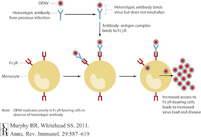
The birth defects and microcephaly accompanying the 2015 - 2016 Zika outbreak in Brazil were well publicised. However, the exact mechanism by which the virus crosses the placenta and infects foetal brain tissue is still being studied. One theory involves the antibody-dependent-enhancement phenomena best known to occur in DENV: where non-neutralising antibodies from previous infections enhance viral replication. Both Zika and Dengue viruses share the Aedes aegypti mosquito vector, and are known to co-circulate in Brazil so it has been posited that pre-existing DENV antibodies may be enhancing viral replication in congenital Zika cases. 

Primary DENV infection  prompts the host adaptive immune response to raise neutralising antibodies for the envelope protein of the virus, inhibiting cellular entry and viral replication. As the immune response matures, antibodies become increasingly specific and cross-reactive to provide long-lasting immunity. These antibodies persist into patient convalescence, and if a secondary Dengue serotype is encountered are thought to promote viral entry into FcγR bearing cells such as monocytes through Fc-receptor-mediated-endocytosis. Secondary Dengue infection thus results in increased viraemia and host inflammatory responses, leading to the increased pathologies of Dengue haemorrhagic fever and shock syndrome.

The findings

From a Thailand cohort containing 16 Dengue-infected children,  researchers gathered plasma and peripheral blood cell samples from acutely infected and convalescent patients. Preliminary testing revealed that plasma from patients convalescing from a secondary Dengue infection bound both DENV and ZIKV, but when neutralising ability against Zika strains HD78788 and PF13 (Africa and French Polynesia respectively) were assessed, the investigators found ZIKV neutralisation to be far less efficient than that of DENV.

If the DENV immunoglobulins are cross-reactive but not neutralising, are they promoting antibody-dependent-enhancement? Moving to a human myeloid cell line U937, which is unreceptive to infection in the absence of ADE. Zika and DENV strains were incubated in DENV-infected serum before addition to the cell line. The resulting infection rate increase in the treatment arm exceeded 100 fold for both Zika viruses, indicating cross-reacting antibodies for DENV were enhancing Zika infection.

Although the Dengue virus exposed plasma could bind Zika viruses, it was not capable of neutralisation and studies showed that presence of DENV antibodies enhanced infection in human cell cultures. a) Capture ELISA showing endpoint titers of DENV-immune plasma in binding ZIKV and DENV.. b) Neutralisation assay of Zika and Dengue viruses using DENV-infected plasma. c) Enhancement of human cell line infection from viruses incubated within DENV-immune plasma. Figures credited to Screaton et al (2016).

The investigators moved onto a panel of human monoclonal antibodies specific for various areas of the DENV envelope protein: one group bound the fusion-loop epitope (FLE), another were specific for the envelope but not FLE, and the remaining 'EDE' third bound intact viral particles with the ability to neutralise infection. Comparing the binding of these anti-Dengue envelope immunoglobulins between the two Zika viruses found the majority of FLE and non-FLE monoclonal antibodies cross-reacted with ZIKV, whilst binding to intact viral particles was variable. When Zika virus was cultured with the first two groups of monoclonal antibodies, infection rates of human myeloid cell lines rose significantly. In a final experiment the team of researchers investigated the EDE group which did not enhance infection, incubating Zika virus with both anti-Dengue patient sera and the various monoclonal antibodies. This study demonstrated that EDE monoclonal antibodies inhibited Zika antibody-dependent-enhancement of infection and protected cell cultures in vivo. 


EDE1 monoclonal human antibodies against DENV are capable of inhibiting antibody-dependent-enhancement of Zika virus in human cell cultures. a) - c) Although the differing monoclonal codes are never truly explained in the text, it is assumed that these refer to differing sets antibodies all binding the EDE1 region. d) Influenza virus was used as a negative control for the inhibition assays. Figures credited to Screaton et al (2016).


Criticism and Caveats

I found myself admiring the simple but elegant experimental design show by this research: each experiment was run with a positive and negative control alongside a series of dilutions. None of the techniques used are new: they're all time-tested virological assays which show that good science can be achieved without the use of the latest gene editing system. I also enjoyed seeing research that used neutralisation and infectivity assays to measure viral load, as opposed to the more common PCR to amplify infected cell RNA. The underlying assumption behind this being increased viral RNA results in increased viral egress, which does logically follow but has not been definitively confirmed for Zika. 


A key assumption of this research is that cohort members were not previously exposured to ZIKV, and the results heavily rely on the fact no reports were made during sampling. Zika virus exposure cannot of course be formally excluded. 
I also feel the research would have benefited from using ZIKV strain from Latin American, as opposed to the African and French Polynesian strains featured but that's slightly pedantic. However, the article lays a solid basis for the ZIKV ADE hypothesis in cell cultures, which will now need to be confirmed in animal models. If readers know of any literature correlating previous DENV exposure with congenital Zika syndrome I'd be very interested to read it. Unfortunately accurate serological testing of this nature is made difficult by the very cross-reactivity shown in this paper. 


Future outlook


This research provides new evidence for the antibody-dependent-enhancement model, but in real world terms what do these results mean? The unfortunate answer is several things, and frankly none of them are very good. These results have the most meaning for Brazil and Latin America where Dengue and Zika are known to co-circulate and share the same vector. There is currently a single Dengue virus vaccine present on the market – Dengvaxia, developed by Sanofi Pasteur - which proved controversial when clinical trials found the treatment arm to increase hospitalisation of children under 9 years old. These results were attributed to the vaccine priming Dengue-naive patients, which resulted in ADE when secondary infections arose. 

This prompted some lively academic discussion, as Scott Halstead of the Dengue Vaccine Initiative called for "further explicit study" in light of ADE evidence, which was not kindly received by members of Sanofi Pasteur. Halstead's reply asking for "a true efficacy study" has not yet been answered and thus questions remain regarding the safety of Dengvaxia. If DENV and ZIKV are found to enhance each other in further animal and human studies, this would be catastrophic for the Dengue vaccine. The described Dengvaxia controversy limited licensing, with use advised for use only in populations with prior DENV seroprevalence of 70%. Attempting to combat these shortfalls, several biotechnology companies are currently developing second generation vaccines and future study of Dengue and Zika interactions will be needed to assess ADE risks between the two viruses.


·       References
1. Dejnirattisai, W., Supasa, P., Wongwiwat, W., Rouvinski, A., Barba-Spaeth, G., Duangchinda, T., Sakuntabhai, A., Cao-Lormeau, V.-M., Malasit, P., Rey, F.A., et al. (2016). Dengue virus sero-cross-reactivity drives antibody-dependent enhancement of infection with zika virus. Nature Immunology 17, 1102–1108..
2. Murphy, B. and Whitehead, S. (2011). Immune Response to Dengue Virus and Prospects for a Vaccine. Annual Review of Immunology, 29(1), pp.587-619.

3. Halstead, S.B., and Russell, P.K. (2016). Protective and immunological behavior of chimeric yellow fever dengue vaccine. Vaccine 34(14), pp.1643–1647.
4.  Hadinegoro, S., Arredondo-García J.L., Guy, B., et al (2016) Answer to the review from Halstead and Russell “Protective and immunological behavior of chimeric yellow fever dengue vaccine”. Vaccine 34(36) pp.4272-4273

5.  Halstead, S.B., and Russell, P.K. (2016). Response to Hadinegoro et al. Vaccine 34(36), pp.4273-4274